Many women with lipoedema are surprised when an assessment includes a look at their veins. After all, lipoedema is a condition of fatty tissue, so why would a vein ultrasound matter? The answer is that lipoedema and venous insufficiency often occur together, and both can contribute to leg pain, heaviness and swelling.
In this article, we explain what venous insufficiency is, what the research says about how often it appears alongside lipoedema, and why a venous assessment can be an important part of understanding your symptoms. We also explain how vein assessment fits into the care pathway at Lipoedema Surgical Solution, including before any surgery is considered, so you know what to expect and why each step is recommended.
What is venous insufficiency?
The veins in your legs carry blood back towards the heart, working against gravity. They rely on small one-way valves to stop blood from flowing backwards. When these valves do not close properly, blood can flow back down the leg and pool, a problem known as venous reflux. Over time, this can lead to chronic venous insufficiency.
Chronic venous disease covers a spectrum. Some people have visible spider veins or varicose veins with few symptoms. Others notice:
- Aching, heaviness or tiredness in the legs, often worse at the end of the day
- Swelling around the ankles and lower legs
- Itching, restless legs or night cramps
- Skin changes, such as discolouration or thickening, in more advanced cases
Several of these symptoms overlap with lipoedema, which is one reason the two conditions can be difficult to separate without a proper assessment.
How are lipoedema and venous insufficiency connected?
Lipoedema is a chronic condition involving an abnormal, usually symmetrical build-up of fatty tissue, most often in the legs and sometimes the arms. Venous disease is a separate condition, but research consistently shows that the two commonly coexist.
The United States consensus guideline on lipoedema lists venous disease among the common conditions that occur alongside it, together with obesity, lymphoedema and joint hypermobility (Herbst et al., 2021). In a 2025 study of 381 women assessed at a Swiss referral centre, chronic venous disease was the most frequently recorded comorbidity, documented in 86.2% of patients (Luta et al., 2025). Because this group was seen at a specialised referral centre, the figure may be higher than in the wider lipoedema population, but it highlights how often veins are relevant.
The reasons for this overlap are still being researched. What matters for patients is practical: if both conditions are present, each may contribute to symptoms, and managing only one may not fully explain or address how your legs feel.
Why are veins checked during a lipoedema assessment?
Lipoedema itself is diagnosed clinically. The 2024 German S2k guideline on lipoedema explains that there is no laboratory or imaging test that confirms it, and that ultrasound has a limited role in diagnosing lipoedema directly. Its main value is in identifying or excluding other causes of swelling, including venous problems (Faerber et al., 2024).
A venous assessment can help to:
- Clarify the cause of swelling. Fluid swelling from venous reflux behaves differently from lipoedema tissue, and knowing what is contributing helps guide management.
- Identify treatable venous disease. If significant reflux or varicose veins are found, treating them may be recommended as part of your plan.
- Support safer planning. If surgery is ever considered, understanding the venous system is an important part of assessing suitability and reducing avoidable risks.
- Provide a baseline. An initial scan gives a reference point for monitoring changes over time.
This whole-of-leg view reflects our phlebology-led approach to lipoedema management. Dr Chris Lekich is trained in phlebology, the area of medicine focused on veins, which is why circulation is considered alongside the fatty tissue itself.
What happens during a venous duplex ultrasound?
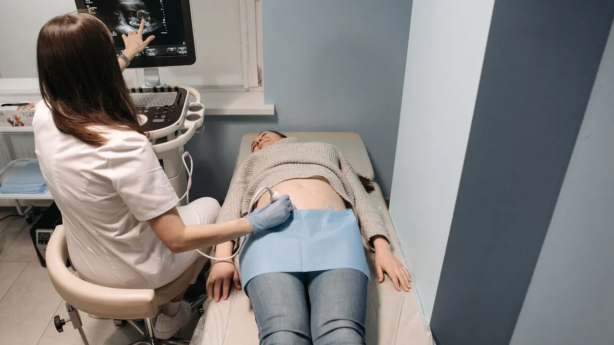
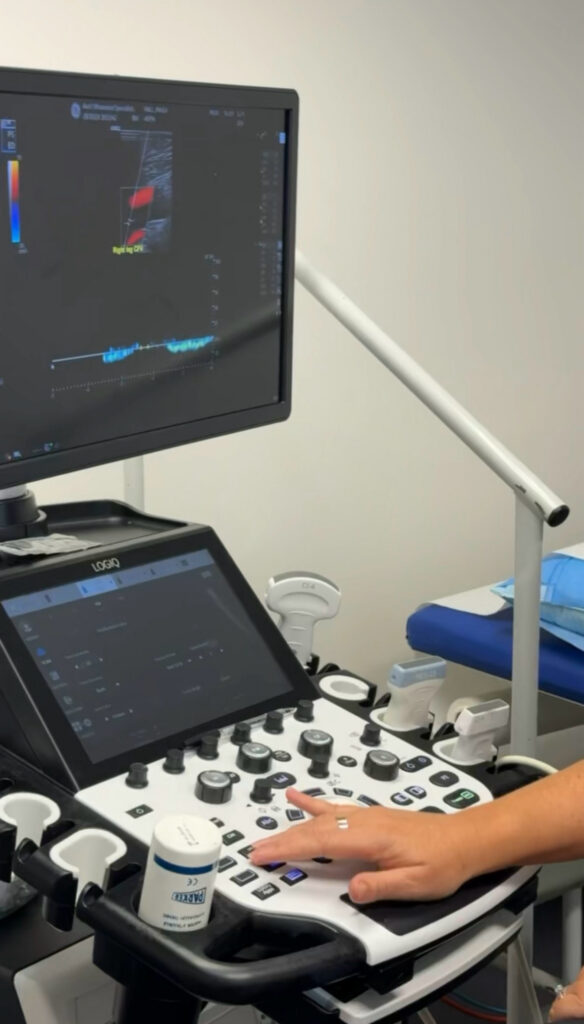
A venous duplex ultrasound is a non-invasive scan that uses sound waves to look at the structure of the veins and how blood is flowing through them. It does not use radiation.
During the scan, you will usually stand or be positioned so the veins fill with blood. A sonographer or doctor moves a probe with gel over the legs, and may gently squeeze the calf or ask you to perform simple manoeuvres to check whether the valves are working. The scan typically looks at both the superficial veins, which lie closer to the skin, and the deep veins.
The results help your doctor understand whether venous reflux is present, how significant it is and whether it may be contributing to your symptoms. Findings are then discussed with you as part of your individual plan.
Lorem ipsum dolor sit amet, consectetur adipiscing elit. Ut elit tellus, luctus nec ullamcorper mattis, pulvinar dapibus leo.
How venous findings can shape your lipoedema plan
Every person’s presentation is different, so venous findings are interpreted in the context of your symptoms, history and goals. In general, they may influence:
Conservative management
Compression is commonly used in both lipoedema and venous disease. Knowing whether venous reflux is present can help guide the type of garment and how it is used. Movement that engages the calf muscle pump, such as walking or water-based exercise, may also support circulation. Our guide to wearing compression for lipoedema has practical tips, and you can read about broader options on our treatment for lipoedema page.
Treating venous disease
How venous disease is treated depends on your individual situation and on what your doctor believes is the most appropriate course of treatment. If you do not wish to have lipoedema surgery, significant venous disease can still be treated in its own right. If surgery is part of your plan, venous treatment may in some circumstances be carried out beforehand, and some venous treatments can be performed at the same time as lipoedema surgery.
Before and after surgery
Surgery is not appropriate for everyone, and many women manage lipoedema conservatively in the long term. For those who do proceed, our onsite ultrasound is used not only in assessment but also after surgery, including for monitoring such as deep vein thrombosis (DVT) surveillance and seroma management. Read more about how surgery is planned on our lipoedema surgery page.
Where can I have a venous assessment with Lipoedema Surgical Solution?
Diagnostic vascular ultrasound is available at our Gold Coast clinic in Miami and at our Brisbane clinic in Spring Hill, where clinically appropriate. Wherever you live, your care with us begins with an online Lipoedema Education Session, followed by a one-on-one telehealth consultation. If you are not able to travel to Miami or Spring Hill, we can refer you for scans locally.
You do not need a GP referral to begin, although your GP can be a valuable part of your care team. To understand the wider assessment process, see our lipoedema diagnosis page.
Frequently asked questions
Is venous insufficiency the same as lipoedema?
No. Venous insufficiency is a problem with blood flow in the veins, caused by valves that do not close properly. Lipoedema is a condition of abnormal fatty tissue. They are different conditions, but they often occur together and can share symptoms such as heaviness and swelling.
Can an ultrasound diagnose lipoedema?
Not on its own. Lipoedema is diagnosed clinically. Ultrasound is mainly used to identify or exclude other causes of swelling, such as venous reflux, which may be contributing to your symptoms.
Does a venous ultrasound hurt?
A venous duplex ultrasound is non-invasive and does not use radiation. Some people feel mild pressure from the probe or when the calf is gently squeezed, particularly if their legs are tender.
Will my veins need treatment before lipoedema surgery?
Not necessarily. It depends on your findings and on what your doctor believes is the right course of treatment for you. Some patients have venous treatment before lipoedema surgery, and some venous treatments can be performed at the same time as surgery. If you do not wish to have surgery, venous disease can still be treated on its own.
Looking after your legs as a whole
Lipoedema and venous insufficiency are separate conditions, but they frequently occur together and can share symptoms such as heaviness, aching and swelling. A clinical assessment, supported by venous ultrasound where appropriate, helps clarify what is contributing to your symptoms and guides a management plan that reflects your whole picture, whether that involves conservative care alone or, for some, surgery in the future.
If you would like to learn more, you are welcome to join one of our lipoedema education sessions, where we explain how assessment, conservative management and treatment options fit together.
Medical disclaimer: This article provides general educational information only and is not a substitute for individual medical advice, diagnosis or treatment. Please speak with your GP or another qualified health practitioner about your own circumstances. Any surgical or invasive procedure carries risks; before proceeding, you should seek a second opinion from an appropriately qualified health practitioner.